Most people know that I work for the University of Auckland within the School of Biological Science as a professional photographer and videographer, and I have done for the last 6 years. A lot of people don't know that, equally important in my job, is my role as a microscopist.
What is a microscopist? I use microscopes, like the one pictured below, to image very small specimens. The specimens could be anything from the micro (that we can't see without magnification), such as whole cells, to the macro (that we can see), such as an insect, plant section, feather etc. I have imaged a very wide range of specimens under the microscope, including genetically modified organisms, plankton, bits of insect, plant sections, crystals, and much, much more.
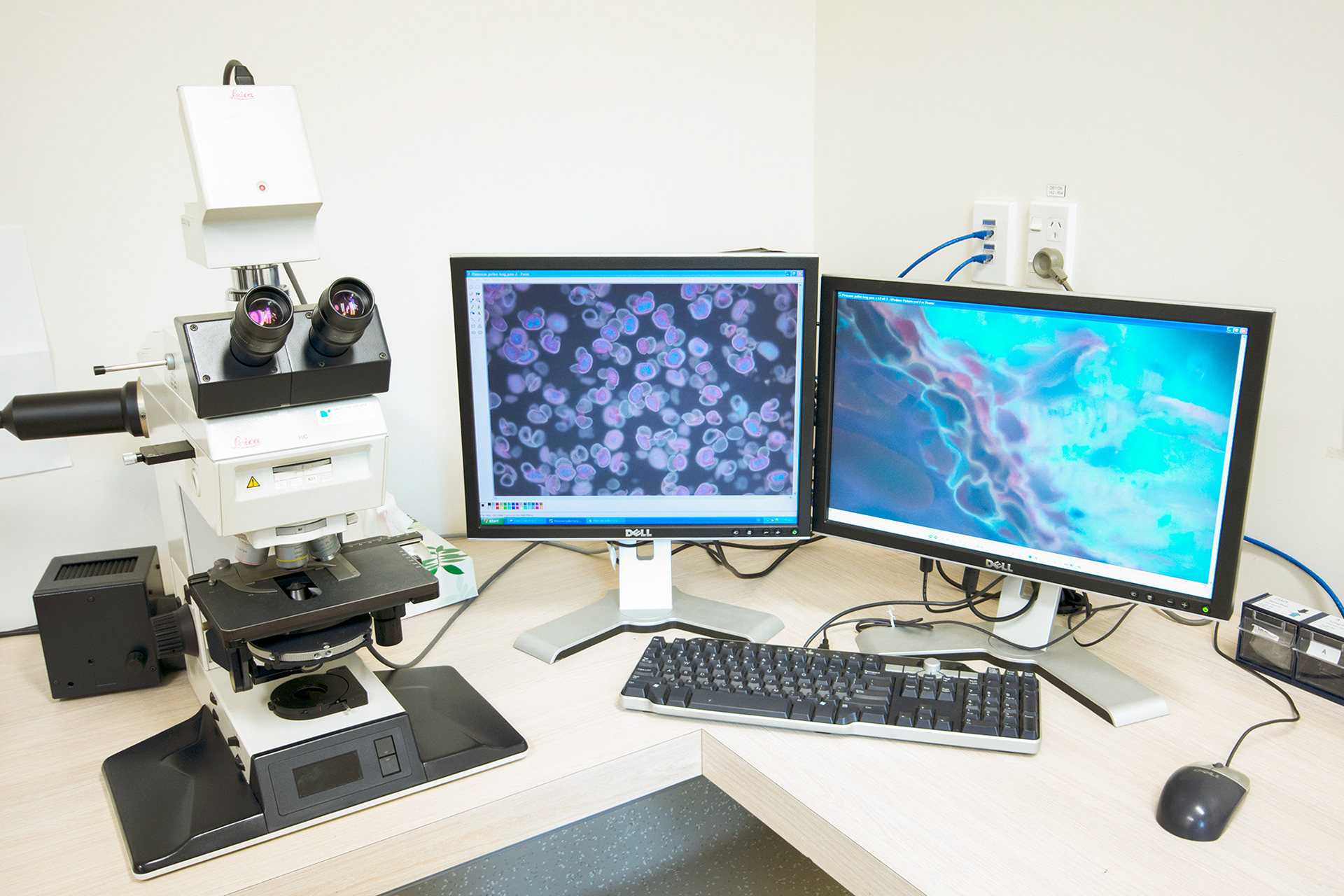
Leica DMRE fluorescence microscope set up at SBS
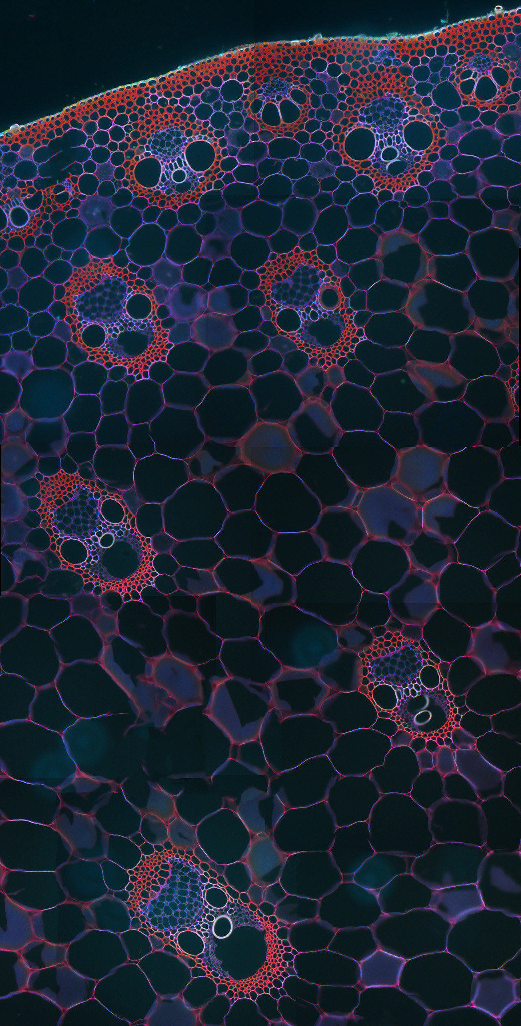
Monocot plant section
The suite of microscopes that I part-manage are what's called light microscopes. They use light to image specimens. This is in contrast to electron microscopes, which fire electrons at specimens to make an image and can subsequently image specimens which are too small for a light microscope to see. This might include viruses, organelles within a cell, and the microstructure of materials. Electron microscopes are huge because they require a vacuum to function and typically take up a whole room. Light microscopes are much more compact and easier to use.
Light microscopes can use and manipulate light in many ways besides using simple white light, or 'bright field'. My favourite is probably polarised light. In photography, we use a single polariser to reduce reflection and enhance colour and contrast. In microscopy, we use 2 polarisers (above and below the sample) to cross-polarise light waves and create a dark background to our image. Many samples appear differently under polarised light (such as the one below), and this appearance can be used to interpret the material qualities of the sample.
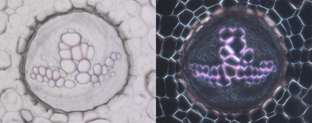
Fern section viewed at 20x magnification under bright field light (left) and polarised light (right)
On our light microscopes, we have the ability to look at samples up to 1,000 times the magnification of the human eye. That's pretty incredible and difficult to imagine. The most common level of magnification is about 200 times the human eye, or more commonly referred to as 20x. That wasn't a typo: 20x is the equivalent of 200 times the human eye. That's because there's a bit of math involved and an extra 10 times magnification between the lens (the 20x lens in this case) and the eyepiece or the camera.
Below is what bacteria look like at 100x (1,000 times the human eye) using a light microscope. If you're a bit disappointed by the lack of detail, you are not alone. You may have heard of the term 'resolution', and you may even have had problems with resolution as a photographer, especially if you shoot macro. Those problems pale in comparison to the constant battle with resolution that microscopists face. The higher we go in magnification, the worse it gets. In addition, a whole new bunch of obstacles present themselves, such as minuscule depth of field, chromatic aberration and diffraction, due to optical limitations and the magnification making these more obvious.
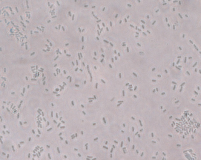
Bacteria under phase contrast at 100x
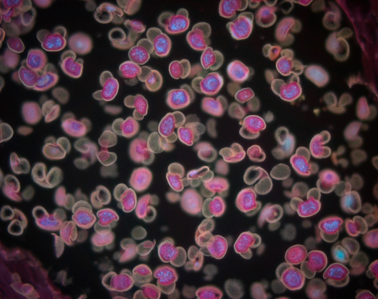
There are multiple ways that a sample needs to be prepared for microscopy, depending on what you want to see. Perhaps most commonly, samples are fixed and sliced into fine sections that light can pass through, typically 1-2 cells thick. These sections are placed onto a glass slide and then stained with dye - to look under normal white light - or introduced to fluorophores - to look under fluorescent light. Fluorophores are basically florescent tags that can specifically target an area of interest in a sample by using antibodies. It's something I did a lot of in my PhD (I was a scientist in a past life). In some cases, commonly in plants, the samples are what's called 'autofluorescent', which means they will fluoresce without dye. The below image of pine cone pollen is an example of this.
Pine cone pollen under fluorescent light, 20x
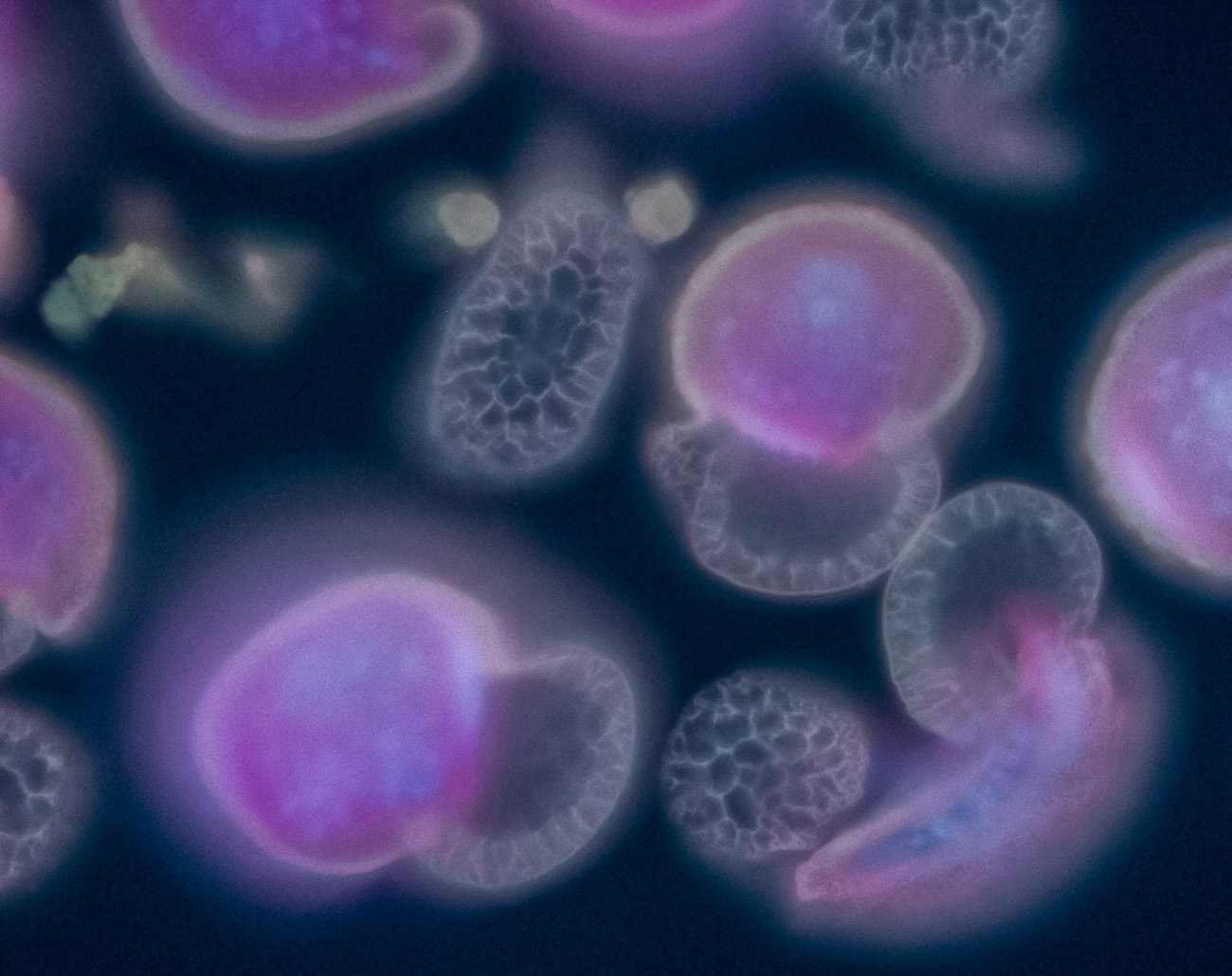
Pine cone pollen under fluorescent light, 100x
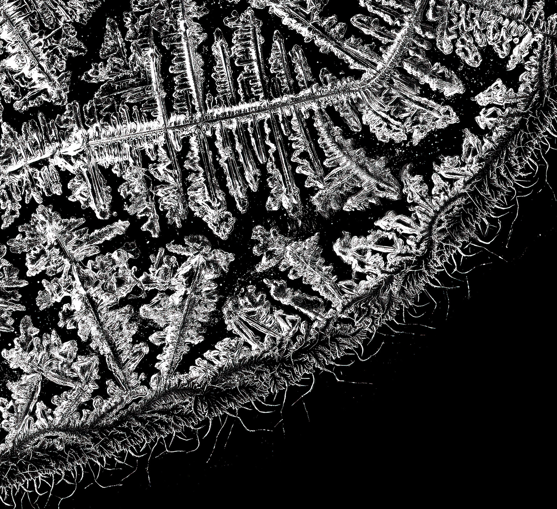
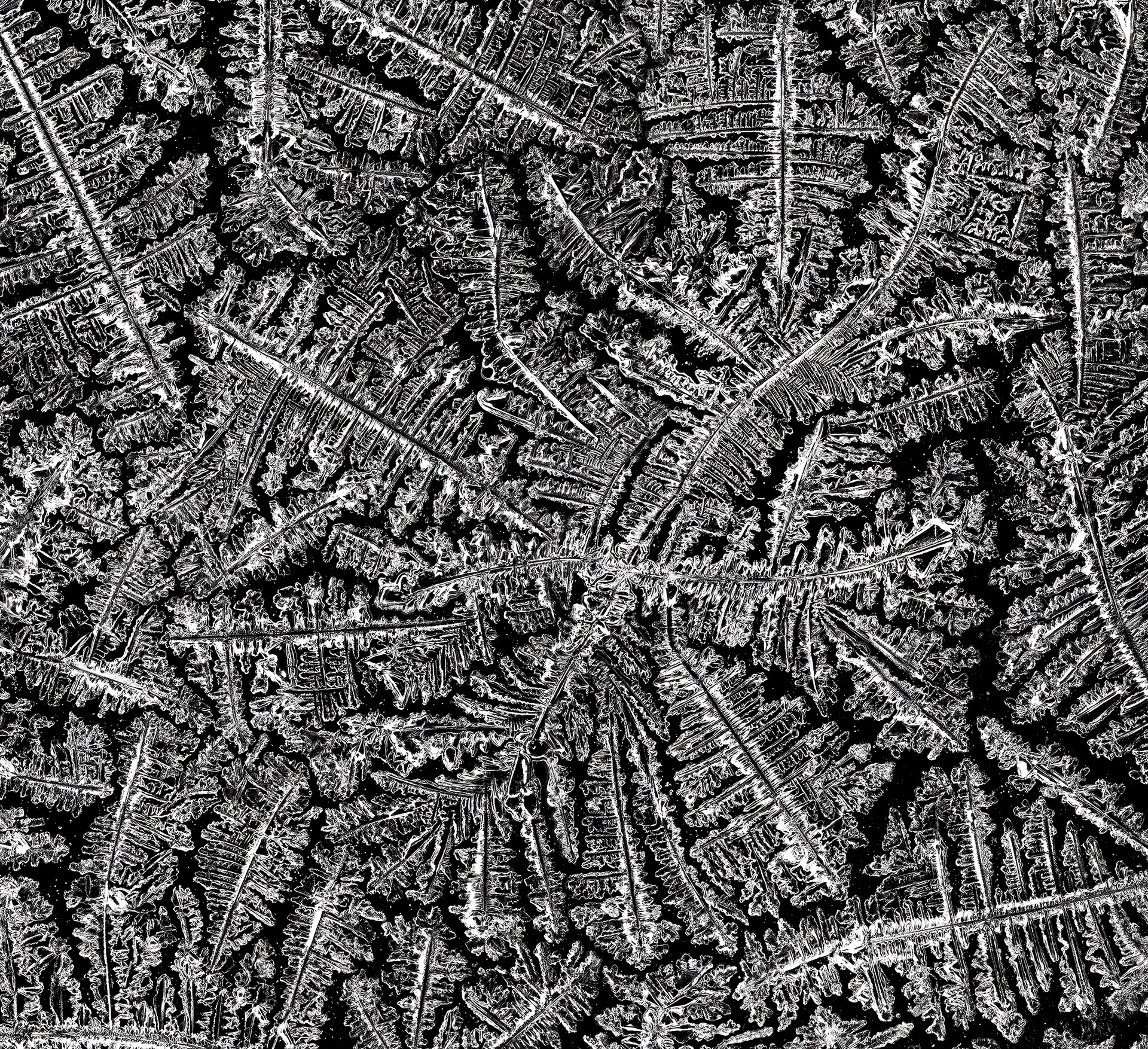
As an artist, I see the potential for images like these as artworks. Indeed, microscopy-related art is not a new concept, and there are competitions for the best works just like in photography. I've done several microscopy-art projects over the years, whether because I had a project in mind, or it was a good way to keep my skills sharp (since I don't do my own research). My latest art project was inspired by coming across magnified images of tears online as a consequence of an unrelated image search. I was immediately transfixed and new I had to try it myself. The below images were taken of one of my tears. After harvesting some of my tears from a good cry, I put a single drop onto a glass slide and then left it alone until it dried. Tears contain all sorts of things, including sodium (hence the salty taste), which can form crystals. I am assuming the crystal formations seen in these images is mostly because of the sodium, which may have been influenced in shape by the other tear contents like enzymes and hormones. I'd like to think that, because I was very sad when I cried, these tears might look different to happy tears, but there isn't a lot of evidence to support this.
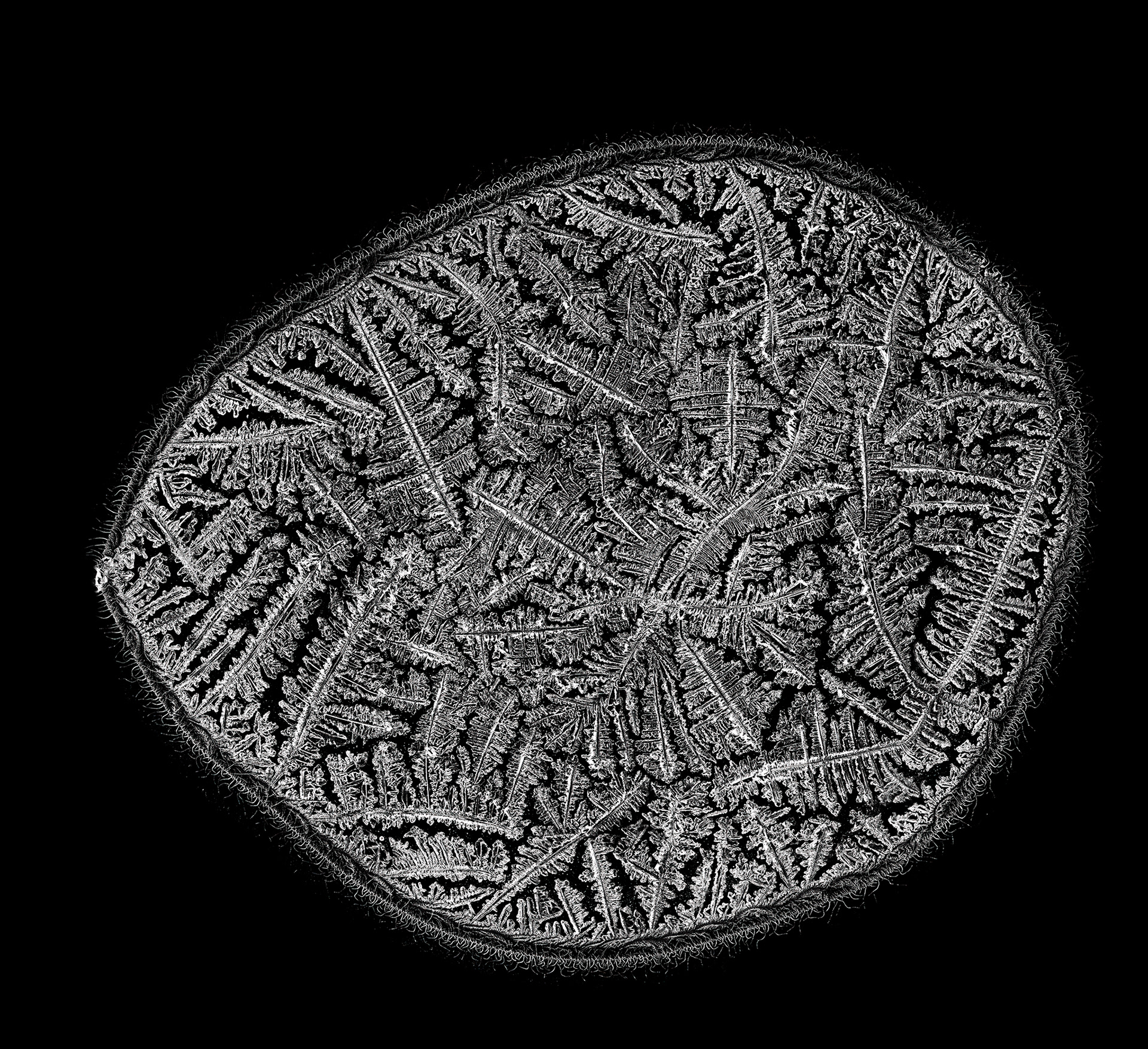
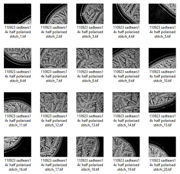
The image above was constructed from 20 individual images captured at 4x under polarised light, collaged together in Photoshop. The main reason I did this was to increase the image size so that I could potentially print it. The cameras on light microscopes are typically around 5MP and capture 72 dpi as standard. Yet another obstacle. Below are some closer crops and underneath those is a screenshot of all the original captures.
Post-processing the composite image involved exposure compensation, increasing contrast, texture and clarity to enhance details, and a lot of sharpening. I then ran the image through Topaz Gigapixel AI to increase it to 300 dpi and enlarge the size. This image can now be printed to a maximum size of A1 without losing detail.
I hope to expand on this project and image tears captured under a variety of situations. Stay tuned!